Optopatcher
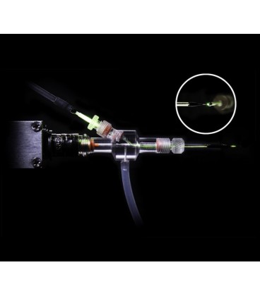
The Optopatcher® provides unmatched accuracy in applying optical stimulation to an in-vivo patch-clamp protocol. The holder houses both an optical fiber and an electrode enabling simultaneous patch-clamp recording and optogenetic activation.
This is a significant advantage in non-slice protocols, such as in-vivo recording from intact animals, whole-cell patch-clamping, LFP recording, and spike/single-unit recording.
The Optopatcher® is available with the most common connectors used on patch clamps: Axon's Threaded Collar, Axopatch and Axoclamp connectors, and standard BNC connectors used by Heka and A-M Systems. It can be ordered to accept capillary glasses with diameters between 1.2 mm and 2.0 mm. For custom diameters, please contact us.
There are two versions of the Optopatcher. The STANDARD configuration of the Optopatcher has a short length internal optic fiber that users can connect their lightsource to via a 1.25mm ceramic sleeve. The CONTINUOUS configuration of the Optopatcher eliminates the internal optic fiber, and uses a single continuous fiber that connects directly to your light source.
The Standard Optopatcher features convenience in use, as the fiber optic can be disconnected to allow removal of the Optopatcher from the rig to ease patch pipette replacement. The Continuous Optopatcher features 10x more light transmission to the target tissue by eliminating the junction between the fiber optic from the light source and the internal optopatcher fiber.
In either style of Optopatcher®, the optical fiber itself needs to be cut to final length for proper fitting within the patch pipette. Previous work by the Optopatcher® developers determined that the fiber end did not need any special tip treatment for effective light transmission. A blunt cut non-polished fiber end could elicit biological responses provided the fiber end was positioned near the end of the pipette.

